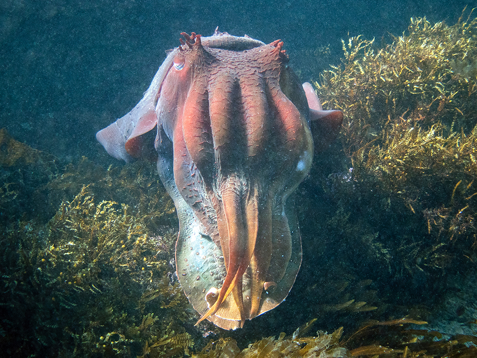
The photo is a front-on view of a very large giant cuttlefish – the biggest I’ve seen in years, perhaps ever. Social media had reports of a large one – “Mr Big” – at Cabbage Tree Bay Aquatic Reserve, north of Sydney (a site central to my book Other Minds). I went looking for him, and others, a few weeks ago.
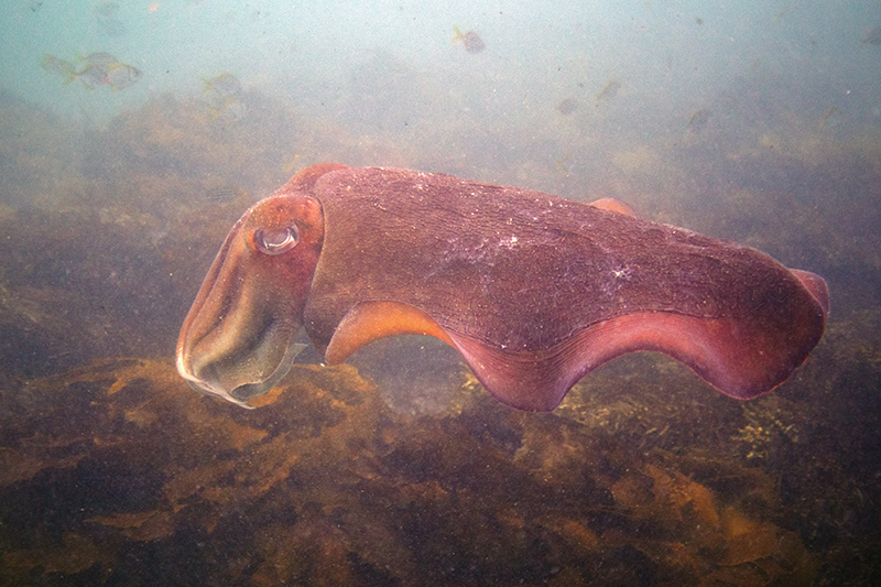
Notes from the snorkel: Conditions were not great – fairly murky. I saw one giant cuttlefish over near the Bower and went on a good roam with him before losing track.
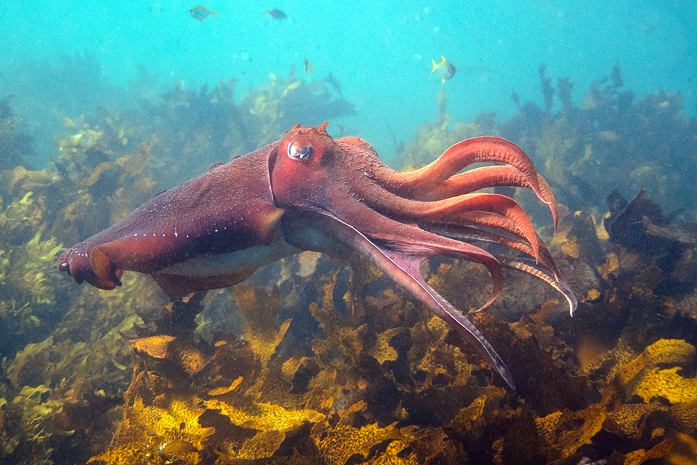
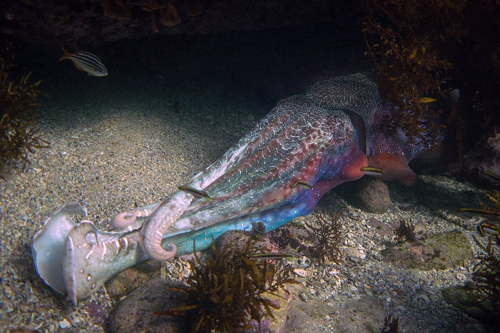
(On instagram, this is Mr Medium.) Then I went up and down the shore, and paused down toward Shelly Beach, when I found a spot where the water was clearer. It seemed a good spot to wait for a while. I saw one male cruising, and he turned out to be a rather determined cuttlefish, medium or medium-small, but quite forthright. Here he is.
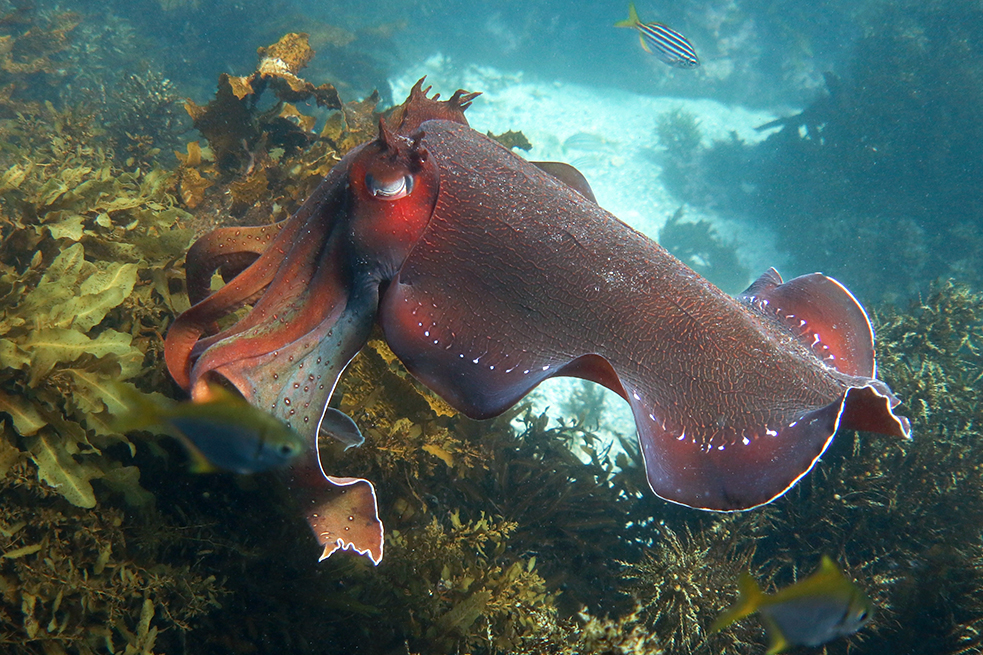 He wandered around, and through his presence and poking under ledges uncovered several others. One of the ones he unearthed was the biggest cuttlefish I’ve seen for a long time – I am pretty sure this was Mr Big.
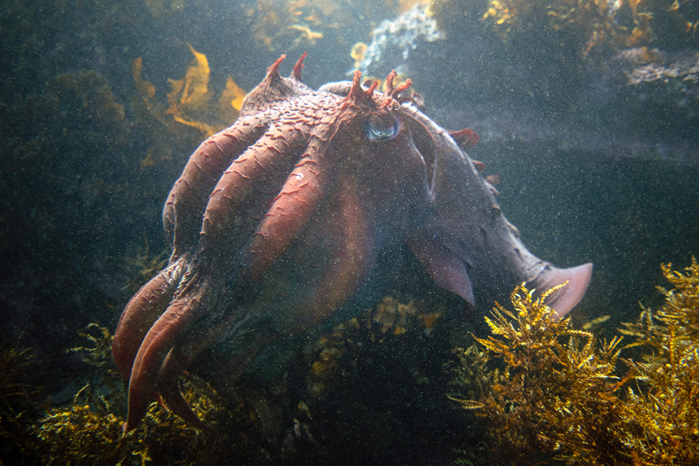
He was at least 1.5 times the length of the medium-sized guy, but probably bigger. He was so big that, unlike other large cuttlefish, he seemed a little ungainly and unwieldy. He wasn’t managing himself in the water as effortlessly as usual, and the ledges and reefs down near the beach didn’t seem to be at the helpful scale that they usually are. He really was too big for the place. He wasn’t senescing; he was pretty much unmarked and very healthy-looking.
I followed him for a while. He didn’t want to engage with the intrepid cuttlefish. The intrepid one was able to make his way around and did not seem intimidated by several much larger cuttlefish. The huge one spent a lot of time beside and half-under a ledge not far from the beach.
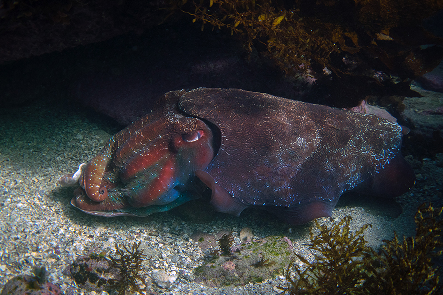
Again, he didn’t seem to fit well underneath it, even when he edged his way in. He did some competitive-yoga displays, but not to anyone I could see.
It was then getting pretty cold, so I said goodbye to him when he was part-under that ledge, and went in. A total of 4-5 cuttlefish that day.
The official size record for a giant cuttlefish, measuring the mantle only (not including the arms at front or the skirt at the back) is 52 cm. Informal reports (Wikipedia etc.) say 100 cm for a large animal with arms out (though not at maximum stretch, I assume, as in the photo above). Mr Big was around that size.
I went back a few days later to look for him again. Now the conditions were awful. No sign of Mr Big, but I did follow one good male cuttlefish as he rocketed through the surge. (Via those water conditions, a photo too bad to post, only link). I am going to go back on Monday [I don’t know when – conditions have been hopeless. Will I make it back before he leaves the scene?]
Postscript: I was able to go back on July 22nd. Lots of surge, but 1-2 good cuttlefish cruising around (not sure whether I saw the same one twice). One of them went to the exact place I’d seen Mr Big, a large ledge not far from Shelly beach (I was glad to locate the place), and poked his head, then whole body, underneath. No sign of Mr Big, and this medium-sized one might have settled in there (or might have emerged a little distance away).
If you write a comment on the post from this front page, the letters will look very small as you type. Write from here to avoid the problem.